HypodontiaHypodontia means that there are fewer than the normal complement of teeth in the dentition. It may be an isolated single tooth missing with no known etiology or it could be associated with a syndrome due to an event occurring before tooth germ formation.
|
|
|
|
The incidence of hypodontia is <1% in the primary dentition incidence and is approximately 2.5-3.5% in the secondary dentition. The secondary teeth most frequently missing are the third molars second premolars maxillary lateral incisors
|
|
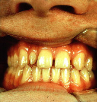
The image above is an example of an abnormality of numbers (congenitally missing permanent maxillary left lateral incisor) and an abnormality of shape. The permanent maxillary right lateral incisor is a peg lateral incisor (courtesy of Drs. Steve Ahing and John Perry, University of Manitoba) |
|
|
Activity not available on mobile devices (description) |
This is an example of a retained primary molar (primary mandibular right second molar) and a single congenitally missing tooth (permanent mandibular right second premolar).
|
|
|
The image on the left is another example of an abnormality of numbers (congenitally missing permanent maxillary right lateral incisor) and an abnormality of shape. The permanent maxillary left lateral incisor is a peg lateral incisor (courtesy of Drs. Steve Ahing and John Perry, University of Manitoba). .
|
|
|
|
|
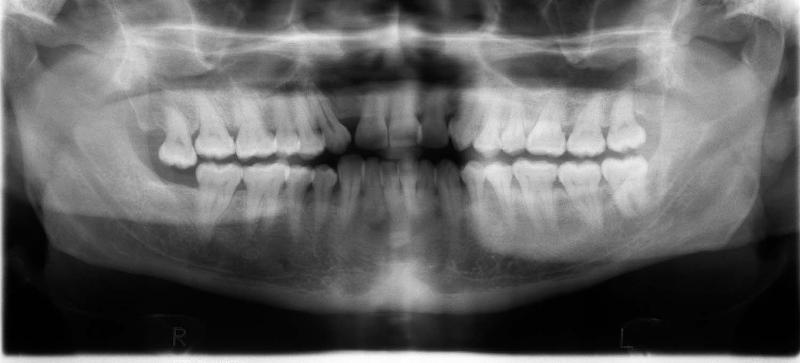
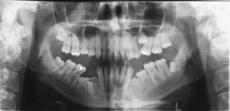
The panoramic radiograph above illustrates a case of a congenitally missing permanent maxillary right lateral incisor. The permanent mandibular right third molar had been previously extracted (courtesy of Dr. BM Cleghorn, Dalhousie University). |
|
|
|
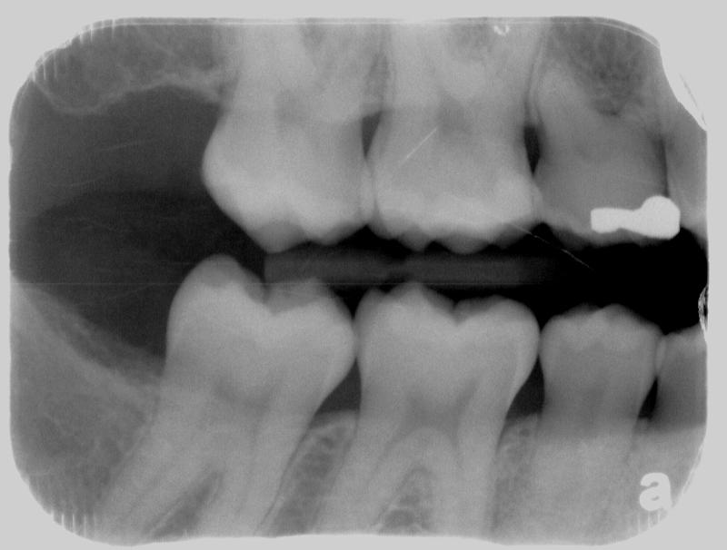
The bitewing radiograph on the left is of the patient's posterior right and illustrates the posterior teeth in quadrants 1 and 4. The primary maxillary right second molar is retained and the permanent maxillary right second premolar is congenitally missing (courtesy of Dr. BM Cleghorn, Dalhousie University). . |
|
|
|
|
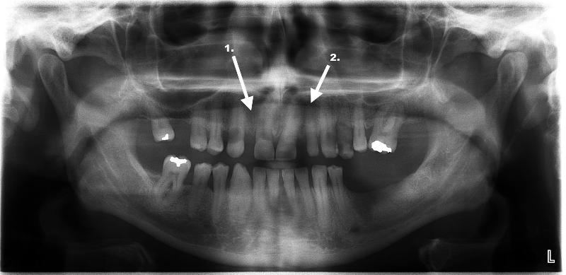
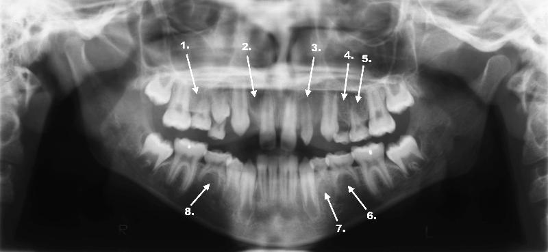
The panoramic radiograph above illustrates a congenitally missing permanent maxillary right lateral incisor (arrow labeled 1.). The arrow labeled 2. is an example of an abnormality in shape as the permanent maxillary left lateral incisor is a peg lateral (courtesy of Dr. BM Cleghorn, Dalhousie University). . |
|
|
|
|
|
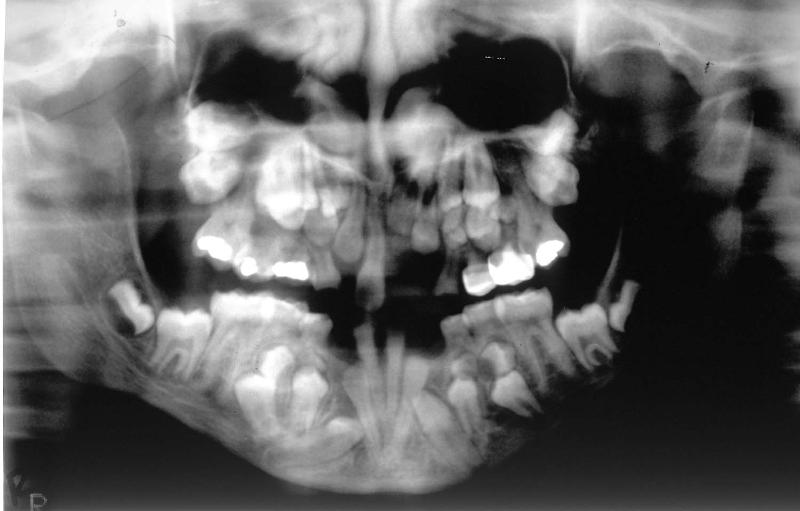
This panoramic radiograph is suggestive of a patient with ectodermal dysplasia. The clinical findings were inconclusive and confirmation of the diagnosis is awaiting genetic testing. Congenitally missing teeth include the permanent maxillary right second premolar, permanent mandibular left first and second premolars and the permanent mandibular right second premolar. Ankylosed primary teeth are present as well as an anomalous permanent maxillary left first premolar. The full case report labeled Case RT is included later in this unit (courtesy of Dr. BM Cleghorn, Dalhousie University). |
|
|
|
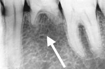
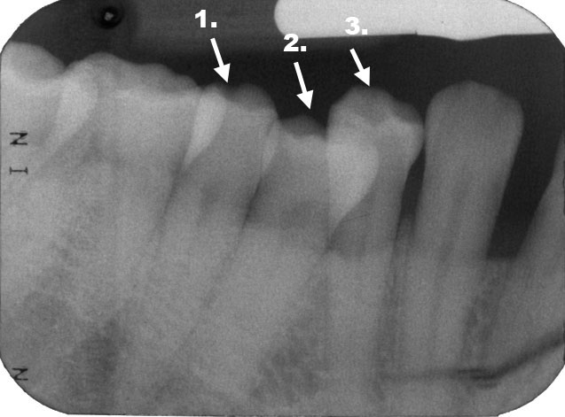
The periapical radiograph on the left illustrates a single congenitally missing tooth. The arrow points to the congenitally missing permanent mandibular left second premolar. Above the arrow is the retained primary mandibular left second molar with partially resorbed roots (courtesy of Drs. Steve Ahing and John Perry, University of Manitoba). |
|
|
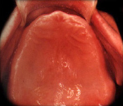
The illustration on the left is an example of induced anodontia. True anodontia is rare. This patient has been rendered edentulous as a result of caries or periodontal disease (courtesy of Drs. Steve Ahing and John Perry, University of Manitoba).
|
|
|
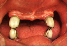
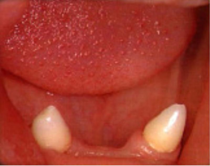
The patient on the left only has four permanent canines remaining. Each of these teeth has been restored with full crowns. This condition is described as partial anodontia (courtesy of Drs. Steve Ahing and John Perry, University of Manitoba). |