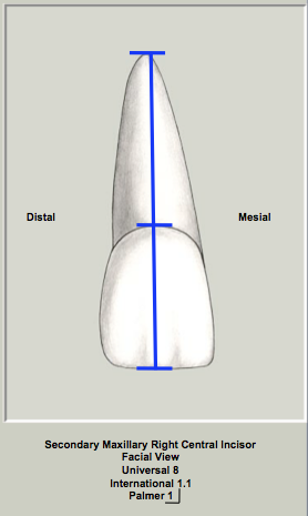
Permanent Maxillary Incisors
Dental Anatomy & Occlusion
Permanent Maxillary Incisors
On completion of the Permanent Maxillary Incisors unit, the student will be able to
1. Correctly use longhand nomenclature for each tooth.
2. Describe each tooth using the following shorthand notations.
Universal or American
Palmer/Zsigmondy
International or FDI
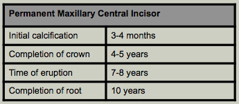
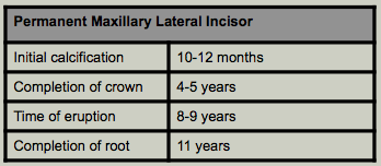
3. Identify the following key tooth development times for this tooth and compare to development times of the other secondary teeth.
Initial calcification
Completion of crown
Time of eruption
Completion of root development
4. Describe the geometrical shape of the crown in this unit from the views listed below and compare to the geometrical shapes of the other secondary teeth.
Facial
Proximal
Incisal or occlusal
5. Describe the location and extent of the facial, lingual, mesial and distal crests of curvature for the tooth in this unit and compare this to the other secondary teeth.
6. Describe the function of each individual tooth and the functions of each class of teeth in each of the primary and secondary dentitions.
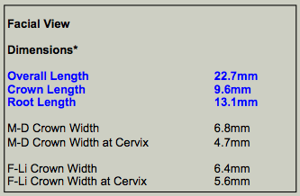
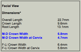
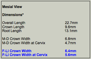
7. Know the average dimensions of the tooth in this chapter and make comparisons to other teeth in the same class, arch and to teeth in different classes, arches and sets.
Overall length
Crown length
Root length
Crown width mesiodistally (at the crest of curvature and at the cervix)
Crown width faciolingually (at the crest of curvature and at the cervix)
8. Describe key anatomical features in each of the following views.
Facial
Lingual
Mesial
Distal
Incisal or occlusal
9. Describe the proximal (mesial and distal) vertical extent of curvature of the cervical lines of the tooth in this chapter and compare to other secondary teeth.
10. List set traits, arch traits, class traits and type traits (as appropriate) for the tooth in this chapter.
11. Describe the location of "normal" maximum intercuspation contacts for the tooth in this chapter.
12. Understand and describe the "normal" root and root canal morphology.
13. Identify variations in crown, root or root canal morphology.
Introduction
|
|
|
Maxillary Incisors
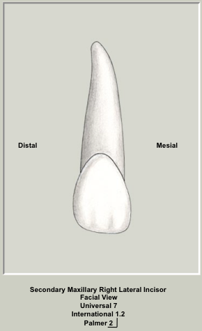
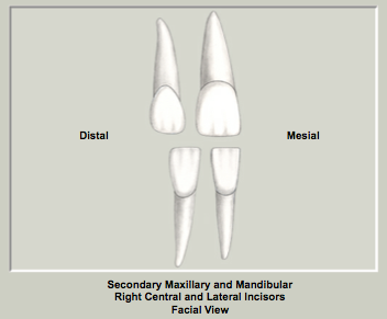
The incisors as a class of teeth have similar geometrical shapes and have similar functions. Each is triangular or wedge-shaped from the proximal view and as the name implies, incisors function by "incising" food. They are also important aesthetically as well. All of the incisors are succedaneous teeth. Each quadrant contains a central and a lateral incisor so there are a total of 8 incisors in each of the primary and secondary dentition. The maxillary incisors are normally single-rooted teeth. The crowns are trapezoidal in shape from the facial view (shorter of the two parallel sides adjacent to the cervix) and triangular from the proximal view. A cross-section of the root at the cervix reveals a conical or triangular shaped root that tapers to the lingual. The maxillary central incisors are located adjacent to the midline and the mesial surfaces of the maxillary right and left central incisors are in contact. Mesial surfaces in contact only occur between the two maxillary central incisors and between the two mandibular central incisors. All other teeth have their mesial surface in contact with the distal surface of the adjacent tooth. The maxillary lateral incisors are located distal (or further from the midline) to the maxillary central incisor in each quadrant. Each of the incisors has an incisal ridge which functions in cutting or shearing food. All other teeth (canines, premolars and molars) have cusps. With wear, the incisal ridge becomes an incisal edge. The secondary (permanent) maxillary central incisors generally erupt after the permanent first molars between the ages of 7 and 8 years. The secondary (permanent) maxillary lateral incisors normally erupt between the ages of 8 and 9 years of age.
|
Tooth Development
|
|
|
|
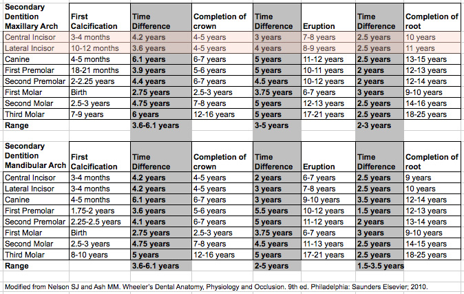
The normal stages of development of each of the primary and secondary teeth are important. The key events are listed in the table. Although there is a range of normal, this knowledge can assist in determining whether your patient's dentition is developing normally or not. Parents will frequently ask about whether their child's dentition is developing normally. In addition, this knowledge is invaluable in determining whether individual teeth are congenitally missing. Aberrations in the timing of development may also provide insight into systemic problems.
Refer to the Tooth Development Table for the Secondary Dentition below to compare the key development times for each of the teeth in the secondary dentition.
|
|
Calcification of all of the teeth in the secondary dentition occurs from birth (permanent maxillary and mandibular first molars) to approximately 10 years of age (maxillary third molar). In this summary table of tooth development, there is a range of 3.6-6.1 years between the time of initial calcification of the crown until the crown is completed.
On average, it takes anywhere from 2-5 years from the time of crown completion until the tooth erupts into the mouth. When teeth erupt, there is approximately 1/2 to 2/3 of the root formed.
After a tooth erupts, further root formation occurs. On average, it takes 1.5-3.5 years for the root to completely form after tooth eruption.
Variations among the timing of development for the permanent teeth are shown in the table to the right.
|
|
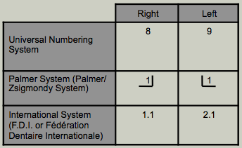
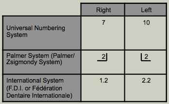
Shorthand Numbering Systems for the Permanent Maxillary Incisors
|
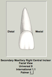
Maxillary Central Incisors
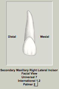
Maxillary Lateral Incisors
|
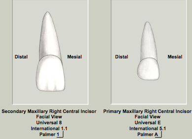
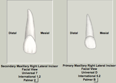
The longhand nomenclature for the maxillary incisors are the permanent maxillary right and left central incisors and the permanent maxillary right and left lateral incisors. The order should be set>arch>side>type for consistency. The three most common shorthand numbering systems for this tooth are described.
The three most common shorthand notations are the F.D.I., Universal and the Palmer systems. Other shorthand notations have been proposed but their use is either limited or currently non-existent. The Universal (American) numbering system is the shorthand system used in the United States. Common to other numbering systems, teeth in the secondary (permanent) dentition are numbered in a clockwise fashion from the maxillary right third molar (1) to the mandibular right third molar (32). This system does not use quadrant numbering but uses consecutive numbers from 1 to 32 to number the permanent teeth. The permanent maxillary right central incisor in the Universal system is tooth number 8 while the permanent maxillary left central incisor is tooth number 9. The permanent maxillary right lateral incisor in the Universal system is tooth number 7 while the permanent maxillary left central incisor is tooth number 10. The numbers 7 (permanent maxillary right lateral incisor), 8 (permanent maxillary right central incisor), 9 (permanent maxillary left central incisor) and 10 (permanent maxillary left lateral incisor) are shorthand numbers unique to the Universal system and no comparable numbers are found in the F.D.I. system.
The Palmer or Palmer/Zsigmondy numbering system uses one of 4 quadrant symbol designations for each of the four quadrants. The maxillary right quadrant is represented by the symbol  . The maxillary left quadrant is represented by the symbol . The maxillary left quadrant is represented by the symbol  . The mandibular left quadrant is represented by the symbol . The mandibular left quadrant is represented by the symbol  . The mandibular right quadrant is represented by the symbol . The mandibular right quadrant is represented by the symbol  . Each quadrant in the secondary (permanent) dentition has eight teeth. The teeth are numbered from 1-8 in each quadrant with 1 representing the central incisor and 8 representing the third molar. . Each quadrant in the secondary (permanent) dentition has eight teeth. The teeth are numbered from 1-8 in each quadrant with 1 representing the central incisor and 8 representing the third molar.
The International (or F.D.I. or Fédération Dentaire Internationale) shorthand system is used throughout the world as the shorthand method of choice except in the United States. This system has two numbers for each tooth. The first number represents the quadrant. Quadrants are numbered in a clockwise manner from the maxillary right to the mandibular right. The quadrants for the secondary dentition in this system are 1 (maxillary right), 2 (maxillary left), 3 (mandibular left) and 4 (mandibular right). The second number in this system represents the secondary tooth from 1 (central incisor) to 8 (third molar). The International system may or may not have a "period" between the quadrant and the tooth. The "official" F.D.I. system does not use a period between the quadrant and the specific tooth. The NDEB (National Dental Examining Board of Canada) utilizes a period between the two numbers. Although not officially correct, this modification does serve to clearly visually differentiate the F.D.I. numbering system from the Universal numbering system. Both forms of the F.D.I. numbering (with and without the period) will be used in this course.
|
Geometric Shape of the Crown
|
Facial & Lingual View
|
|
|
|
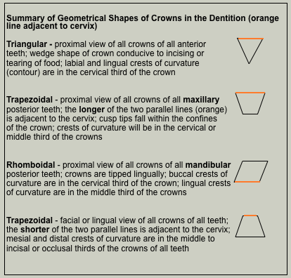
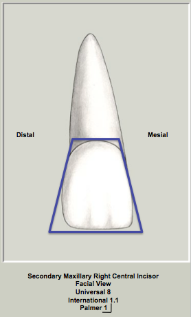
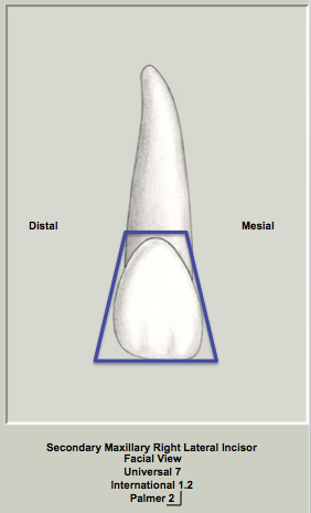
The geometrical shape of the crowns from the facial and lingual view is trapezoidal. The shorter of the two parallel sides is adjacent to the cervix. This geometrical shape is the same for the facial (and lingual) views of all crowns in both the secondary and primary dentitions. This basic geometrical shape means that all crowns taper towards the cervix. The mesial and distal crests of curvature vary amongst the teeth in the dentition, but this basic shape indicates that the mesial and distal crests of curvature of all teeth are either in the middle to incisal (occlusal) thirds of the crowns.
|
Mesial & Distal (Proximal) View Crown
|
|
|
|
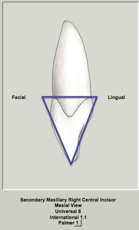
The geometrical shape of the crowns of the maxillary central and lateral incisors from the mesial and distal view is triangular. The base of the triangle is adjacent to the cervix (or CEJ or cementoenamel junction). This geometrical shape is the same for the proximal (mesial and distal) views of all anterior teeth in both dentitions.
|
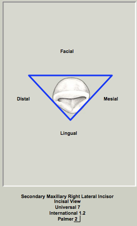
Incisal View
|
|
|
|
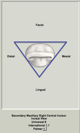
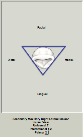
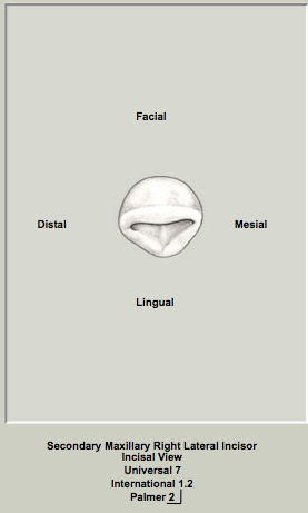
The geometrical shape of the crowns of the maxillary central and lateral incisors from the incisal view is triangular. All of the incisors (maxillary and mandibular) have this basic geometrical shape. The base of the triangle is positioned labially and the apex of the triangle is lingual due to the lingual taper of the crown.
This differs from the canines, which are diamond-shaped from this view, due to their highly developed middle labial lobe.
Dimensionally, the crown of the permanent maxillary central incisor is larger mesiodistally and labiolingually than the permanent maxillary lateral incisor.
|
Crest of Curvature (Contour)
|
Facial View
|
|
|
|
The crest of curvature (contour) is defined as the widest area of the crown of the tooth when viewed from the facial (or lingual) or proximal (mesial or distal). Each tooth has a distinct location for the crest of curvature and it is based on the crown divided into thirds.
The crest of curvature is synonymous with the contact area only when teeth are in perfect alignment. When teeth are not in perfect alignment, the contact area will not correspond to the crest of curvature.
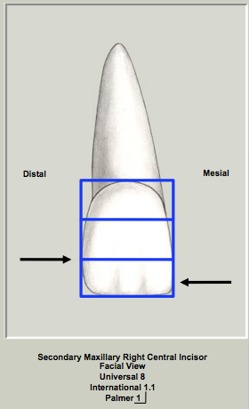
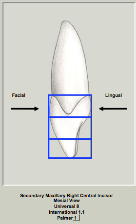
Maxillary Central Incisor
The mesial crest of curvature (contour) is located in the incisal third of the crown virtually at the mesioincisal corner. The distal crest of curvature (contour) is located at the junction of the middle and incisal third of the crown.
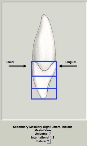
Maxillary Lateral Incisor
The mesial crest of curvature (contour) is located at the junction of the middle and incisal third of the crown. The distal crest of curvature (contour) is located in the middle third of the crown.
|
Proximal View
|
|
|
|
|
|
Maxillary Central Incisor
The labial and lingual crests of curvature for the permanent maxillary central incisor is located in the cervical third of the crown.
Maxillary Lateral Incisor
The labial and lingual crests of curvature for the permanent maxillary lateral incisor is also located in the cervical third of the crown. The locations of the labial and lingual crest of curvature (contour) are consistent with the triangular geometrical shape of the crown in this view.
|
Extent of Crest of Curvature
|
|
|
|
|
|
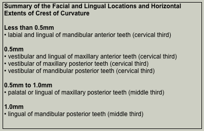
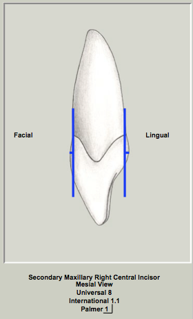
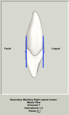
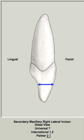
This is a horizontal measurement of how "bulbous" the tooth is. The maxillary anterior teeth have a labial and lingual extent of the crest of curvature of 0.5mm.
|
Summary of Geometrical Shapes of Crowns & Extent of Crests of Curvature
Function
The function of the maxillary incisors is consistent with their shape. The maxillary incisors are wedge-shaped in the proximal view and each incisor has a sharp incisal ridge so their function is to incise or cut food. This wedge-shape in the proximal view is shared with the mandibular incisors and the maxillary and mandibular canines. Therefore they also function in incising or cutting food.
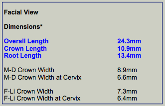
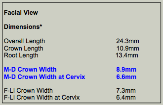
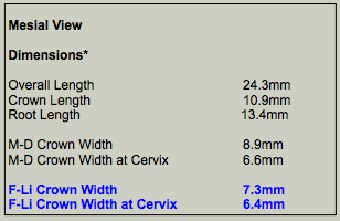
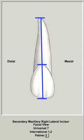
Dimensions of the Maxillary Central Incisor
Dimensions of the Maxillary Lateral Incisor
External Morphology Maxillary Central Incisor
|
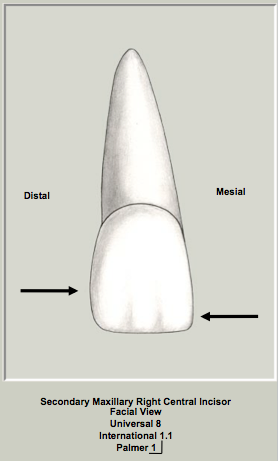
Facial view
|
|
|
Crests of Curvature
The mesial and distal crests of curvatures are at different levels as the mesial surface contacts the adjacent central incisor and the distal surface is in contact with the lateral incisor. The mesial crest of curvature is in the the incisal third of the crown and the distal crest of curvature is located at the junction of the middle and incisal third of the crown. The outline of the crown is a series of curves. The mesial crown outline is slightly convex while the distal outline of the crown is more convex. The crown has an incisal ridge which is slightly convex from mesial to distal. Two basic crown shapes exist; one is wide M-D at the cervix (square crown form) and the other is narrow M-D at the cervix (tapered crown form). In the facial view, the cervical line is convex.
|
|
|
Incisal Ridge
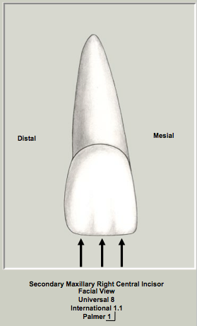
There is evidence of three mamelons visible in the unworn (newly erupted) tooth (see arrows). With wear, the incisal ridge becomes an incisal edge. The incisal ridge is slightly curved mesiodistally so that the crown has its greatest length in the centre of the incisal ridge of the tooth.
Labial Surface of Crown
Three convexities are visible on the labial surface that demarcate the three labial lobes.
Root
The root is cone-shaped and convex on its labial surface. There is a conical shape to the root which has greater taper in the apical half compared to the cervical half of the root. Overall the root is usually about 2 or 3mm longer than the crown. There is often a curvature of the apex to the mesial or distal and this curvature is variable. The apex is usually slightly blunt.
|
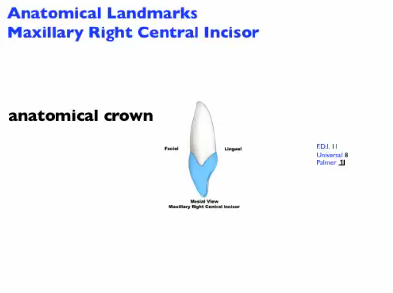
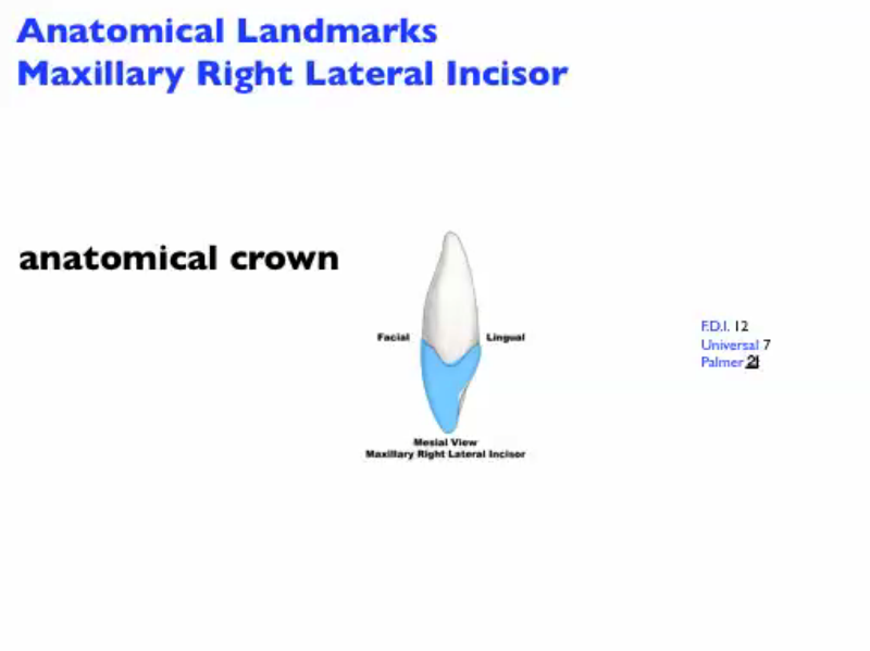
Anatomical Landmarks of the Maxillary Right Central Incisor - Facial View
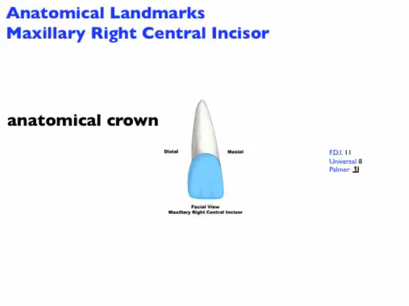
Click on this hyperlink to view the anatomical landmarks in the facial view.
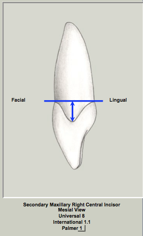
Mesial View
|
|
|
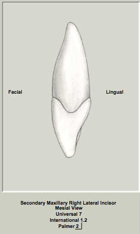
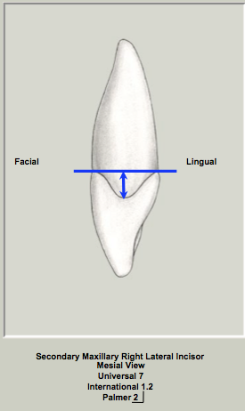
The crown is triangular in shape or wedge-shaped like all of the anterior teeth from this view with the apex of the triangle at the incisal ridge and the base of the triangle at the cervix of the tooth.
Facial Outline of Crown
The crest of curvature labially is located in the cervical third of the crown. The labial outline of the crown is convex especially in the cervical third. The crown is less convex from the crest of curvature to the incisal ridge.
Lingual Outline of Crown
The cingulum is convex in the cervical third of the crown. From the cingulum to the incisal ridge, the lingual crown surface is concave. The lingual surface again becomes convex approaching the incisal ridge.
Cervical line
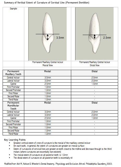
Note the greater curvature of the cervical line on the mesial surface of the maxillary central incisor compared to the maxillary lateral incisor. At approximately 3.5mm, the curvature of the cervical line on the mesial surface of the permanent maxillary central incisor exhibits the greatest vertical extent of any surface of any tooth in the dentition.
Mesial Surface of the Crown
The crown is generally convex in this view.
Incisal ridge
The location of the incisal ridges of the permanent maxillary incisors are described as being on a line bisecting the root or slightly labial to a line bisecting the root. This should be compared to the mandibular incisors which have the incisal ridge lingual to a line bisecting the root. This feature that allows differentiation of teeth in a class between the arches is called an arch trait.
Root
The root is conical in form and the majority of the root taper occurs in the apical half of the root.
|
Anatomical Landmarks of the Maxillary Right Central Incisor - Mesial View
Click on this hyperlink to view the anatomical landmarks in the mesial view.
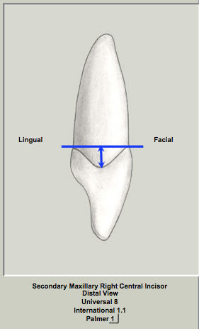
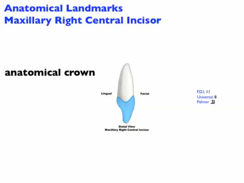
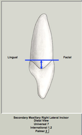
Distal View
|
|
|
|
The crown is triangular in shape or wedge-shaped like all of the anterior teeth from this view. The distal aspect of the maxillary central incisor is similar to the mesial view. Differences between the two proximal views will be pointed out.
Facial Outline of Crown
No differences from the mesial view are present. The crown is convex from cervix to the incisal ridge.
Lingual Outline of Crown
The lingual outline is similar to the mesial outline.
Cervical line
The distal cervical line curves approximately 2.5mm which is 1mm less than the mesial curvature of the cervical line.
|
|
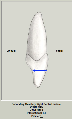
|
The crown appears wider labiolingually from this view as more of the labial surface of the crown can be seen in this view.
Incisal Ridge
No differences from the mesial view are noted.
Root
The root has a similar conical shape comparable to the mesial view.
|
Anatomical Landmarks of the Maxillary Right Central Incisor - Distal View
Click on this hyperlink to view the anatomical landmarks in the distal view.
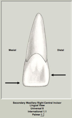
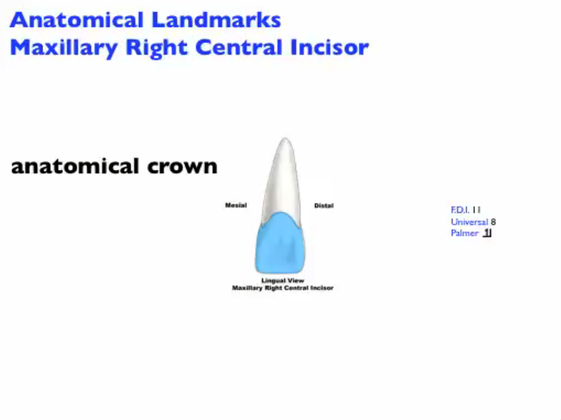
Lingual View
|
|
|
Both the crown and root taper towards the lingual. The lingual aspect of crown is a series of convexities (mesial marginal ridge, distal marginal ridge, cingulum and the lingual aspect of the incisal ridge called the linguoincisal ridge) and a concavity (lingual fossa). The cervical line is convex.
Mesial Outline of Crown
The mesial outline is slightly convex (much less so compared to the distal outline) and the mesial crest of curvature is at the mesioincisal corner of the crown.
Distal Outline of Crown
The distal outline of the crown is convex and the distal crest of curvature is at the junction of the middle and incisal thirds of the crown.
Incisal Ridge
The incisal ridge from the lingual view is convex and the linguo-incisal ridge is visible.
Lingual Surface of Crown
The cingulum is present in the cervical third of the crown. The lingual anatomy of crown is characterized by the convex cingulum, mesial and distal marginal ridges and incisal ridge. These convexities of the lingual surface border the concave lingual fossa which is a large, centrally located region. Developmental grooves can be present on the lingual surface of the crown (cingulum to lingual fossa).
Root
The root is convex on its lingual surface and tapers lingually consistent with its conical or triangular shape in cross-section.
|
|
|
|
Anatomical Landmarks of the Maxillary Right Central Incisor - Lingual View
Click on this hyperlink to view the anatomical landmarks in the lingual view.
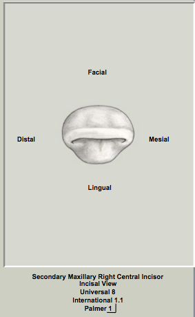
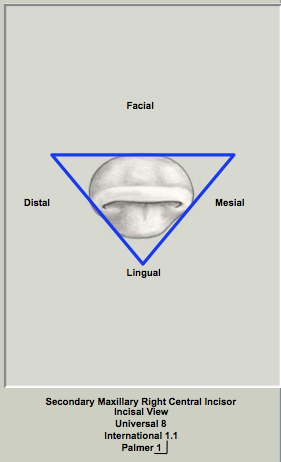
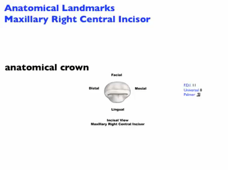
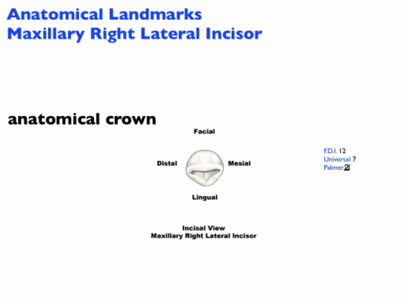
Incisal View
|
|
|
|
The shape of the crown in the incisal view tends to be triangular like all of the incisors and the crown tapers towards the lingual.
Facial Outline of Crown
The crown is evenly convex in this view at the crest of curvature but flattens out from the crest of curvature to the incisal ridge.
Lingual Outline of Crown
The cingulum is prominent in this view and is wide mesiodistally.
Mesial Surface of the Crown
The crown is generally convex.
Distal Surface of the Crown
The crown surface is generally convex.
|
|
|
Incisal Ridge
In this view, the incisal ridge is centred labiolingually or slightly labial to the line. The difference between the labiolingual and mesiodistal widths should be noted. The mesiodistal width is greater than the labiolingual dimension by approximately 1.5mm.
Root
The root is not visible in this view.
|
Anatomical Landmarks of the Maxillary Right Central Incisor - Incisal View
Click on this hyperlink to view the anatomical landmarks in the incisal view.
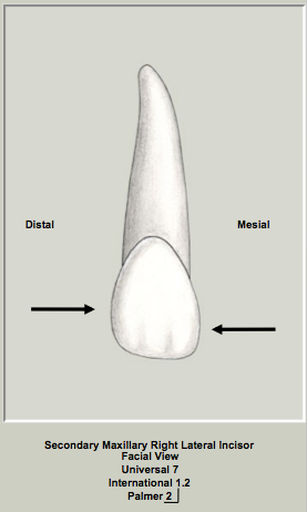
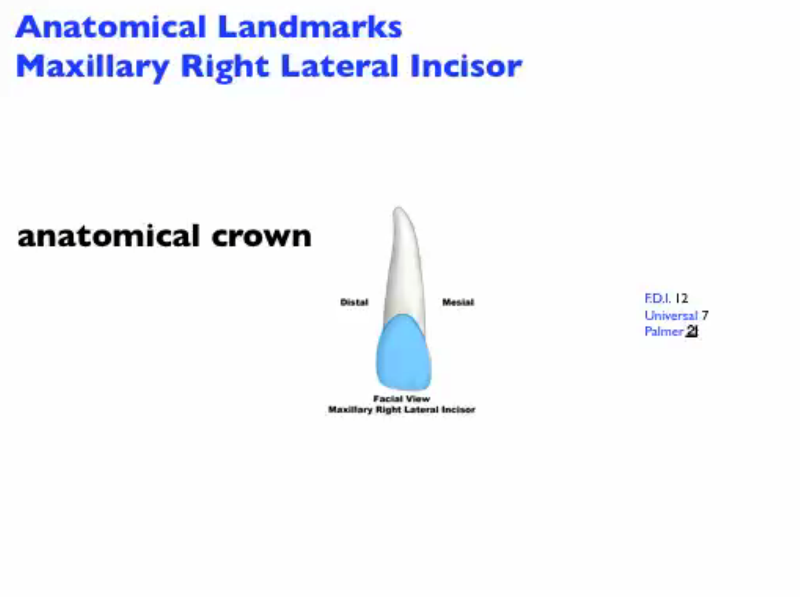
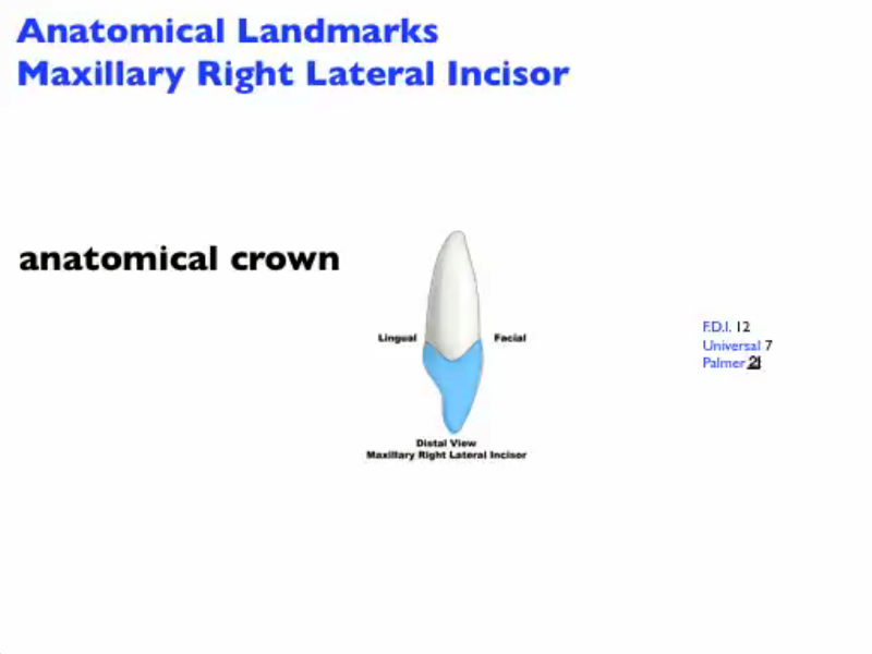
External Morphology Maxillary Lateral Incisor
|
Facial view
|
|
|
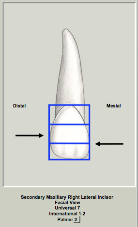
The mesial and distal crests of curvatures are at different levels as the mesial surface contacts the distal of the central incisor and the distal surface is in contact with the mesial surface of the canine. The outline of the crown is a series of curves. The distal outline of crown is more convex than the mesial outline and both of these outlines are more convex than the maxillary central incisor. The crown has an incisal ridge which is convex from mesial to distal and is more convex compared to the maxillary central incisor. Two basic crown shapes exist; one is wide M-D at the cervix (square crown form) and the other is narrow M-D at the cervix (tapered crown form). The cervical line is convex in this view.
Mesial Outline of Crown
The mesial outline of the crown is convex. The mesial crest of curvature is at the junction of the incisal and middle third of the crown.
Distal Outline of Crown
The distal outline is more convex than the mesial outline of the crown and the distal crest of curvature is in the middle third of the crown.
Incisal Ridge
With wear, the incisal ridge becomes an incisal edge. The incisal ridge is curved mesiodistally.
Labial Surface of Crown
Three convexities are visible on the labial surface that demarcate the three labial lobes. There is generally more cervical taper mesiodistally compared to the maxillary central incisor.
Root
The root is cone-shaped and convex on its labial surface. There is a conical shape to the root which has greater taper in the apical half compared to the cervical half of the root. Overall, the root is usually about 1.5X longer than the crown. There is often a curvature of the apex to the distal but this is variable.
|
Anatomical Landmarks of the Maxillary Right Lateral Incisor - Facial View
Click on this hyperlink to view the anatomical landmarks in the facial view.
Mesial View
|
|
|
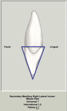
The crown is triangular in shape or wedge-shaped like all of the anterior teeth from this view with the apex of the triangle at the incisal ridge and the base of the triangle at the cervix of the tooth. The C:R (crown:root) ratio is less compared to the maxillary central incisor (the root of the maxillary lateral incisor is longer in relation to the crown on the maxillary lateral incisor).
Facial Outline of Crown
The crest of curvature labially is located in the cervical third of the crown. The labial outline of the crown is convex especially in the cervical third. The crown is convex from the cervix to the incisal ridge. From the crest of curvature to the incisal ridge, the maxillary lateral incisor is more convex than the maxillary central incisor.
Lingual Outline of Crown
The cingulum is convex in the cervical third of the crown. From the cervix to the incisal ridge, the lingual crown surface is concave. The lingual surface again becomes convex approaching the incisal ridge.
|
|
|
Cervical line
The cervical line curves approximately 3.0mm towards the incisal ridge (vertical measurement of the curvature). This curvature is slightly less than that found on the mesial of the permanent maxillary central incisor (3.5mm).
Mesial Surface of the Crown
The crown is generally convex.
Incisal ridge
The incisal ridge is normally on a line bisecting the root.
Root
The root is conical in form in cross-section. The majority of the root taper occurs in the apical half of the root.
|
Anatomical Landmarks of the Maxillary Right Lateral Incisor - Mesial View
Click on this hyperlink to view the anatomical landmarks in the mesial view.
Distal View
|
|
|
The crown is triangular in shape or wedge-shaped like all of the anterior teeth from this view. The distal aspect of the maxillary lateral incisor is similar to the mesial view. Differences between the two views will be pointed out.
Facial Outline of Crown
No differences from the mesial view are noted.
Lingual Outline of Crown
This view is similar to the mesial outline.
Cervical line
The distal cervical line curves approximately 2.0mm which is 1mm less than the mesial curvature of the cervical line.
|
|
|
Distal Surface of the Crown
The crown appears wider labiolingually from this view as more of the labial surface of the crown can be seen in this view.
Incisal Ridge
No differences from the mesial view are noted.
Root
• similar conical cross-sectional shape as the mesial view
• the root tapers to a blunt apex with the majority of the taper occurring in the apical half of the root
|
Anatomical Landmarks of the Maxillary Right Lateral Incisor - Distal View
Click on this hyperlink to view the anatomical landmarks in the distal view.
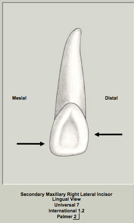
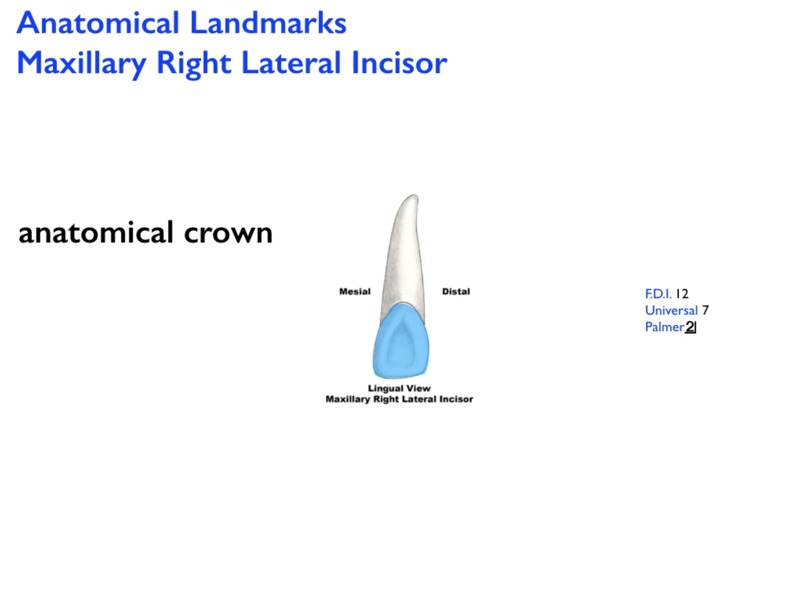
Lingual View
|
|
|
|
The crown and root both taper towards the lingual. The lingual aspect of crown is a series of convexities (mesial marginal ridge, distal marginal ridge, cingulum and the lingual aspect of the incisal ridge called the linguo-incisal ridge) and a concavity (lingual fossa). The cervical line is convex.
Mesial Outline of Crown
The mesial outline is convex. The mesial crest of curvature is at the junction of the middle and incisal thirds of the crown.
Distal Outline of Crown
The distal outline is more convex than the mesial outline and the distal crest of curvature is in the middle third of the crown.
Incisal Ridge
The incisal ridge is convex and curved mesiodistally. The linguo-incisal ridge is visible in this view.
Lingual Surface of Crown
The cingulum is present in the cervical third of the crown. The lingual anatomy of crown is characterized by the convex cingulum, mesial and distal marginal ridges and incisal ridge. The convexities of the lingual surface border the lingual fossa which is a large, centrally located region. Developmental pits and grooves can be present on the lingual surface of the crown (cingulum to lingual fossa) and can extend onto the root as a radicular groove.
Root
The root is convex on its lingual surface and tapers lingually consistent with its conical shape in cross-section.
|
Anatomical Landmarks of the Maxillary Right Lateral Incisor - Lingual View
Click on this hyperlink to view the anatomical landmarks in the lingual view.
Incisal View
|
|
|
|
The shape of the crown in the incisal view tends to be triangular like all of the incisors or it can have a diamond-shape like the canines. The crown tapers towards the lingual.
Facial Outline of Crown
The crown is evenly convex in this view and tends to be more convex than the maxillary central incisor.
Lingual Outline of Crown
The cingulum is prominent in this view but is more narrow than the maxillary central incisor or the canine.
Mesial Surface of the Crown
The crown is generally convex.
Distal Surface of the Crown
The crown surface is generally convex.
|
|
|
Incisal Ridge
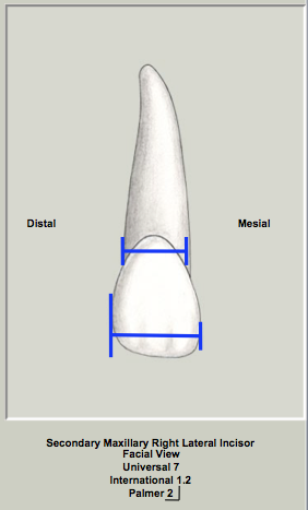
In this view, the incisal ridge is centred labiolingually or is slightly labial to a line bisecting the root. The difference between the labiolingual and mesiodistal widths should be noted as the difference is less (6.8mm mesiodistally vs 6.4mm labiolingually) than the maxillary central incisor. The mesiodistal width is greater than the labiolingual dimension only by 0.4mm.
Root
The root is not visible in this view.
|
Anatomical Landmarks of the Maxillary Right Lateral Incisor - Incisal View
Click on this hyperlink to view the anatomical landmarks in the incisal view.
|
Roots
|
|
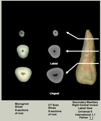
Maxillary Central Incisor
|
The root of the maxillary central incisor is not visible from the incisal view. However, a cross-section of the central incisor at the CEJ reveals a conical or triangular root form. The root tends to be triangular at the cervix and more conical in the middle and apical portions of the root. The root tapers towards the lingual so the base of the triangle is labial and the apex of the triangle is oriented lingually.
|
Maxillary Lateral Incisor
|
The root of the maxillary lateral incisor is not visible from the incisal view. However, a cross-section of the lateral incisor at the CEJ reveals a conical or ovoid root form. The root tapers towards the lingual.
|
Summary of the Vertical Extent of the Curvature of the Cervical Line for the Permanent Dentition
Set Traits
|
|
|
Maxillary Central Incisors
Set Traits are characteristics that differentiate teeth in the primary and secondary dentitions. Traits of the secondary tooth will be listed that are different from their primary counterparts.
The crowns of the secondary maxillary central incisors have a greater crown length (cervicoincisal) than crown width (primary maxillary central incisors are wider mesiodistally compared to their crown length).
The mesial and distal crests of curvature of the secondary maxillary central incisors are at different locations. The mesial crest of curvature is in the incisal third of the crown approaching the mesioincisal corner of the crown and the distal crest of curvature is located at the junction of the middle and incisal third of the crown. The primary central incisors have mesial and distal crests of curvature in the incisal third of the crown with the distal slightly more cervical in its position. They are virtually in the same position mesially and distally.
There is less cervical constriction mesiodistally and labiolingually in all secondary teeth compared to their primary counterparts.
The crown:root ratio is less for all primary teeth compared to their secondary counterparts. Primary teeth have much longer roots in comparison to their crowns compared to secondary teeth.
All of these characteristics apply to both the maxillary central and lateral incisors.
|
|
|
Maxillary Lateral Incisors
The crowns of the permanent maxillary lateral incisors have a greater crown length (cervicoincisal) than crown width as do the deciduous maxillary lateral incisors but the deciduous maxillary lateral incisors differ in proportions and appear to have a shorter, wider crown in comparison.
The mesial and distal crests of curvature of the permanent maxillary lateral incisors are at different locations- mesial at the junction of the middle and incisal third of the crown; distal in the middle third of the crown (deciduous maxillary lateral incisors have mesial and distal crests of curvature in the incisal third of the crown with the distal slightly more cervical in its position).
There is less cervical constriction mesiodistally and labiolingually in all permanent teeth compared to their deciduous counterparts.
The crown:root ratio is less for all deciduous teeth compared to their permanent counterparts (deciduous teeth have much longer roots in comparison to their crowns compared to permanent teeth).
|
Arch Traits
|
|
|
Arch traits are characteristics that differentiate the maxillary and mandibular teeth in the same class.
The incisal ridge in maxillary incisors is centred labiolingually while mandibular incisors have the incisal ridge lingual to a line bisecting the root.
In the maxillary arch, the permanent maxillary central incisor is larger than the permanent maxillary lateral incisor while the opposite is true in the mandibular arch.
The permanent maxillary incisors have a deeper, more well-defined lingual fossa compared to the flatter, less developed lingual fossa of the mandibular incisors.
The permanent maxillary central and lateral incisors are wider mesiodistally than their mandibular counterparts.
The cingula of the permanent maxillary incisors are more prominent compared to the mandibular incisors.
A cross-section of the root of the permanent maxillary incisors is conical or triangular while the root of the mandibular incisors is hour-glass shaped (wide labiolingually with mesial and distal root depressions).
The crowns of permanent maxillary incisors are wider mesiodistally than labiolingually while the opposite is true of the mandibular incisors.
|
Class Traits
|
|
|
Class traits are characteristics that are unique to a specific class of teeth.
All incisors have an incisal ridge.
Incisors have 3 mamelons present on the incisal ridge upon eruption.
All incisors are wedge-shaped or triangular in the incisal view.
All incisors have a single lingual fossa.
|
Type Traits
|
|
|
Maxillary Central Incisor
Type traits are the most specific type of traits that make a tooth unique.
The maxillary central incisor is the widest mesiodistally of all anterior teeth.
The mesiodistal dimension of the crown is closer to the cervicoincisal dimension when compared to any of the other incisors. In other words, the crown is more square in shape rather than being narrow and long as are the other incisors.
The vertical curvature of the cervical line on the mesial surface of the permanent maxillary central incisor is the greatest of all teeth in either dentition (3.5mm).
|
|
|
Maxillary Lateral Incisor
The permanent maxillary lateral incisor exhibits more variation in its anatomical form than does any other tooth except for the third molars.
Variations include
• peg lateral incisors (deficient mesial and distal lobes)
• caniniform (appears canine-like with a small cusp)
• congenitally missing
• toed lateral (prominent mesioincisal corner with cervical constriction)
• small central incisor (more square crown with less cervical constriction)
• radicular groove
The permanent maxillary lateral incisor has the least difference between its mesiodistal and labiolingual dimensions of any of the anterior teeth (0.5mm)
The maxillary lateral incisor has the highest incidence of radicular grooves of all of the incisors (approximately 3.0% - Source: Pecora JD, da Cruz Filho AM. Study of the Incidence of Radicular Grooves in Maxillary Incisors. Braz Dent J 1992;3(1):11-16.)
Lingual pits are more commonly on the maxillary lateral incisor than any of the other incisors.
|
Maximum Intercuspation Contacts
|
|
Contact position and size
|
|
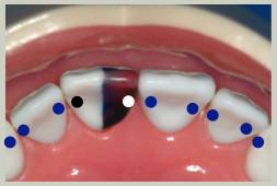
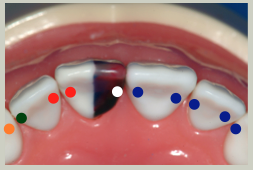
Maxillary Central Incisors
|
The incisal ridge of mandibular central incisor (white dot) is in contact with the mesial marginal ridge of the maxillary central incisor and the incisal ridge of mandibular lateral incisor (black dot) is in contact with the distal marginal ridge of the maxillary central incisor.
|
Maxillary Lateral Incisors
|
The incisal ridge of mandibular lateral incisor (red dots) is in contact with the mesial marginal ridge of the maxillary lateral incisor and the distal marginal ridge of the maxillary central incisor.
The mesial cusp ridge of the mandibular canine (green dot) is in contact with the distal marginal ridge of the maxillary lateral incisor and the distal cusp ridge of the mandibular canine (orange dot) is in contact with the mesial marginal ridge of the maxillary canine.
|
VR Models
Maxillary right central incisor
Click and hold down the cursor to rotate and turn the crown (or click on the 3D model and use the arrow buttons).
Models for the VR movies provided courtesy of Viade Products, Inc.
Maxillary right lateral incisor
Click and hold down the cursor to rotate and turn the crown (or click on the 3D model and use the arrow buttons).
Models for the VR movies provided courtesy of Viade Products, Inc.
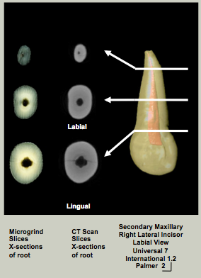
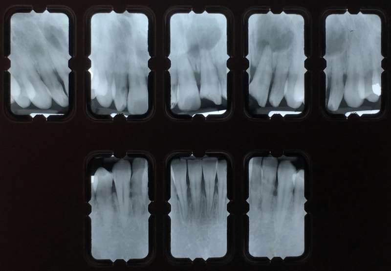
Summary of Root & Root Canal Morphology
|
Maxillary Central Incisors
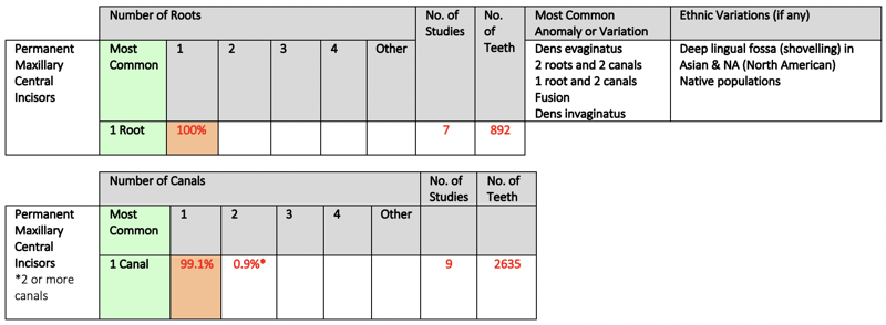
The maxillary central incisor is normally a single-rooted tooth with a single root canal system.
|
|
Maxillary Lateral Incisors
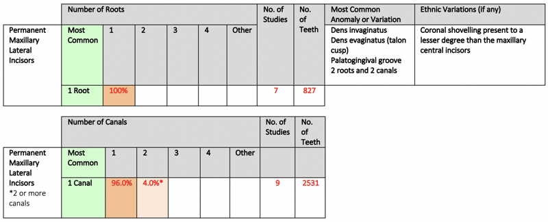
The maxillary lateral incisor is normally a single-rooted tooth with a single root canal system.
|
|
|
Anomalies
|
Maxillary Central Incisors
Talon cusps (dens evaginatus) on both of the permanent maxillary central incisors.
|
Maxillary Lateral Incisors
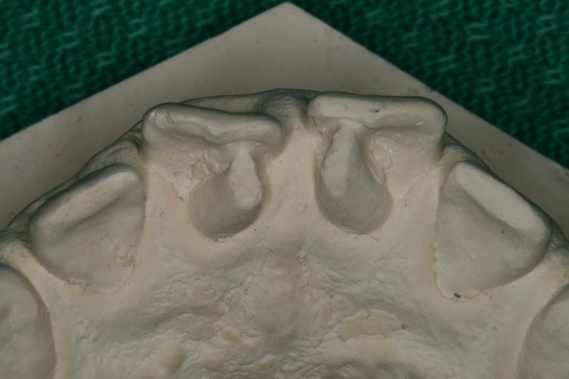
 Dens invaginatus in both of the permanent maxillary lateral incisors. Dens invaginatus in both of the permanent maxillary lateral incisors.
|

References
Ash M, Nelson S. Wheeler's Dental Anatomy, Physiology and Occlusion. 8th ed. Philadelphia: Saunders; 2003.
Bjorndal, AM, Henderson, WG, Skidmore, AE and Kellner, FH. Anatomic measurements of human teeth extracted from males between the ages of 17 and 21 years. Oral Surg 1974;38:791-803.
Black G. Descriptive Anatomy of the Teeth. 4th ed. Philadelphia: SS White Dental Manufacturing Company; 1902.
Brown P, Herbranson E. Dental Anatomy & 3D Tooth Atlas Version 6.4. 6th ed. Portola Valley: eHuman; 2010.
Dewey M. Dental anatomy. St. Louis: C.V. Mosby; 1916.
Diamond, M. Dental Anatomy. New York: The Macmillan Co.; 1929.
Fuller JL, Denehy GE, Schulein TM. Concise dental anatomy and morphology. 4th ed. Iowa City, IA: University of Iowa College of Dentistry; 2001.
Jordan RE, Abrams L. Kraus' dental anatomy and occlusion. St. Louis: Mosby Year Book; 1992.
Linek, HA. Tooth Carving Manual. New York: Columbia Dentoform Co.; 1949.
Nelson SJ, Ash MM. Wheeler's dental anatomy, physiology, and occlusion. 9th ed. St. Louis, Mo.: Saunders/Elsevier; 2010.
Pecora JD, da Cruz Filho AM. Study of the incidence of radicular grooves in maxillary incisors. Braz Dent J 1992: 3: 11-6.
Renner RP. An introduction to dental anatomy and esthetics. Chicago: Quintessence Pub. Co.; 1985.
Scheid RC, Woelfel JB. Woelfel's dental anatomy. 8th ed. Philadelphia: Wolters Kluwer/Lippincott Williams & Wilkins Health; 2010.
Shillingburg, HT, Kaplan, MJ and Grace CS. Tooth dimensions - a comparative study. J South Calif Dent Assoc 1972;40:830-839. Woelfel, JB and Scheid, RC. Dental Anatomy. 6th ed. Philadelphia: Lippincott Williams & Wilkins; 2002.